
In the history of medicine, there are very few diagnostic procedures which have had the impact of magnetic resonance imaging, more commonly known as MRI. The MRI scanners use magnetic fields, radio waves and field gradients to form images of the anatomy, and they are capable of revealing the physiological properties of the body, including disease and injuries.
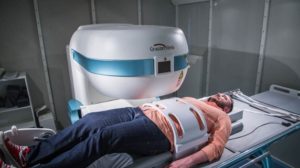
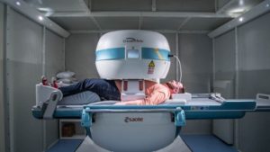
The MRI technology of the past has only allowed for a patient to be scanned in a lying position. This position does not always provide accurate insight for diagnosis because it does not show how a patient’s body weight affects their spine. The weight-bearing MRI system allows the physician to image patients in the position they experience discomfort which helps lead to a more accurate diagnosis.
The MRI process was patented and introduced to physicians in the mid 1970’s, and one of the early adopters of this technology was the Texas Back Institute (TBI). I was one of the three founders of TBI, and I would like to share some insights about the value of an MRI scan for spine diagnosis and how we have taken this technology to a new level.
The MRI Has Become a Valuable Diagnostic Tool
 The MRI scan is a unique way of looking at our patients’ spines. It allows us to look at the bones and nerves to determine if there is any compression on the nerves in the spinal canal. The unique thing about the MRI scan is that it involves no X-rays and therefore no radiation. Most of us know about the negative effects of getting too many X-rays, CT scans and the like. This works off of magnetic resonance, and to date, there have been no reports of harm to the human body from this type of scan.
But how does an MRI work, and how are the images obtained?
Basically, a magnetic field is passed through the body. This interacts with the magnetic field found around each of our human cells. Then with computer enhancement, we are able to see unbelievable pictures of the spine and its individual nerve roots that exit each level to innervate our muscles and give us our sensations. It’s really an incredible technology.
“Science is marching forward, and we love being on the cutting edge of it!”
A New MRI Technology
The Texas Back Institute has a rich history of pioneering new medical technology, and the organization has one of the first MRIs designed for both load-bearing and non-load bearing scans to see their impact on the joints, including the spine.
It’s a more compact MRI scan. Plus, it is much cheaper than the traditional MRI scan and it works off a lower magnetic field. Just as we see advancements in other fields, such as electronics, cell phones or computers, the developers of this new MRI, with very low magnetic fields, are able to get excellent images.
 In the past, our patients who have had previous surgeries which required metal implants, such as screws, rods and cages, were not able to get reliable MRIs because the implants would cause interference with the magnetic field and prevent it from producing clear images. With this low magnetic field scan, these patients are now able to benefit from this technology, and they have developed special software that allows us to see better images of the nerves and the bones despite the hardware.
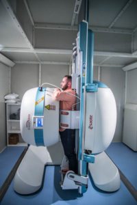
This new MRI has another important advancement. It has the ability to take upright images.
The reason this is important is that in many spinal abnormalities, patients don’t hurt when they are lying down. They say, “Doctor, I hurt when I’m up on my feet, when I stand too long or when I’m walking.” Now, we have the ability to take an MRI scan when the patient is in a standing position as well.
This is what we call being in a “loaded” position, where the weight of the body is being transmitted through the spine and disc, and it may actually cause a bulging of the disc. This can cause a pinching of the nerves and may also cause instability. These conditions are not going to be visible when the patient is lying flat on their back.
I’ve had numerous patients who have normal MRI scans while they were lying on their backs, and when you stand them up for regular, plain X-rays, we see that they have abnormal motion.
 The most common reason for taking an MRI scan is to help diagnose the reason a patient might have persistent back and leg pain and is not responding to conservative treatment,” Dr. Guyer said. “Most patients might have a little back sprain that will get better within a few weeks. However, if they begin to have pain or symptoms which are not responsive over the initial two or three weeks of treatment, then the physician would probably order an MRI scan. Or, if the patient reports having weakness, numbness or other neurologic symptoms, then we would probably go to an MRI scan earlier.”
What’s Next?
As one of the three physicians who formed the Texas Back Institute more than 30-years ago, I have experienced tremendous changes in the practice of medicine and spine care specifically. In the beginning, did we ever think that innovations such as this new MRI would be such an important part of the practice? No.
When we first started using MRI scans, many years ago, the initial images weren’t very good and we all said, “Well, this is not going to be good for spines.” Well, we were so wrong! Now the quality and resolution is so much better.
However, I do think there will be advances in the future that will give us total body scans. It’s not going to stop here. Science is marching forward, and we love being on the cutting-edge of it!
If you have a patient with persistent back pain and feel this new MRI might be a valuable diagnostic tool, contact Dr. Richard Guyer or any other of the spine specialists at the Texas Back Institute in Plano, Texas by calling 800.247.2225.
If you or a family member is experiencing persistent back pain, contact your family physician or the Texas Back Institute at 800.247.2225.
For more information visit www.texasback.com.


Recent Comments